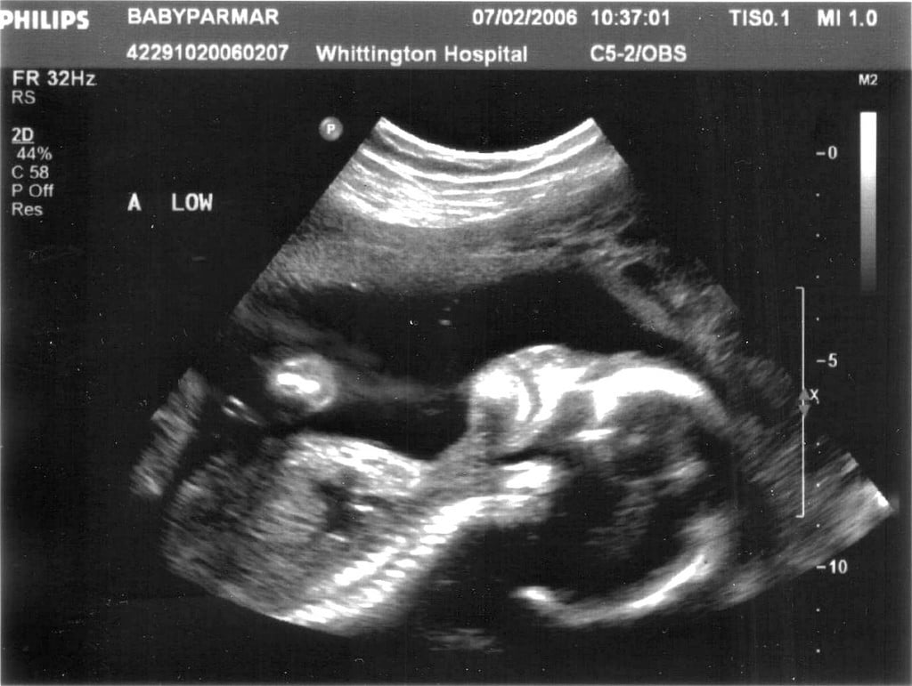
Identify the indications for an abdominal ultrasound. This activity highlights the role of the interprofessional team in the care of patients undergoing this procedure. This activity reviews the use of ultrasound in the detection of many abdominal pathologies, with a particular focus on indications, contraindications, and technique involved in performing a transabdominal ultrasound. Ultrasound is portable, cost-effective and does not require radiation or contrast. The concept includes the assessment or evaluation of the quality of care identification of problems or shortcomings in the delivery of care designing activities to overcome these deficiencies and follow-up monitoring to ensure effectiveness of corrective steps.Ultrasound is an imaging modality that has been in clinical use for approximately 50 years, though its utility and efficacy have dramatically improved since it was first introduced. Determines the depth of the ultrasound beam quality Quality Activities and programs intended to assure or improve the quality of care in either a defined medical setting or a program. Differentiation of 2 objects close to each other, parallel to the beam. Axial Axial Computed Tomography (CT) definition:. Image definition or sharpness of the image generated can be characterized in terms of: Cine images: captured during real-time scanning. Transverse: perpendicular to the sagittal Sagittal Computed Tomography (CT) plane. 
Sagittal Sagittal Computed Tomography (CT) (or longitudinal): along the long axis of the structure being evaluated.The diagram shows that as the ultrasound wave beam (blue horizontal bar) penetrates the tissues, a percentage is reflected back (left arrows) toward the transducer while another continues to go deeper into the tissues (right arrow), losing some energy to the parenchyma as it goes. The CPU processes the electrical signals into images that can be seen on the monitor.Lower amplitudes are assigned shades closer to black.Higher amplitudes are assigned shades closer to white.The signals are assigned a shade of gray depending on the amplitude of the echo produced by the tissue after interacting with the piezoelectric crystals. The sound waves are turned into electrical signals and then amplified in the console.Absorbed energy from the beam is later released as heat Heat Inflammation.Digestion and Absorption of the emitted beam. The amplitude of the echoes depends on the degree of energy absorption Absorption Absorption involves the uptake of nutrient molecules and their transfer from the lumen of the GI tract across the enterocytes and into the interstitial space, where they can be taken up in the venous or lymphatic circulation.As the beam travels, it is reflected by structures in the tissues (or echoes) back to the transducer, with some energy being absorbed by the tissues.Sound waves penetrate the tissues in the form of a beam.
 Sound waves are emitted by the transducer. The main principle behind ultrasound imaging is the transmission and reflection of sound waves through the tissues. License: Public Domain Generation of images with ultrasound Image by Lecturio.Īn ultrasound machine and different probes Image: “Photos of a sonography system and typical transducers.” by Kieran Maher. However, this comes at the cost of image resolution. Note that decreasing the frequency increases the depth to which the ultrasound wave travels. Activation of M-mode M-Mode Imaging of the Heart and Great Vessels and Doppler. Allows for the manipulation of the images coming from the transducer. Central processing unit (CPU): processes electrical signals to generate an image. Frequency is inversely related to wavelength and depth of tissue penetration Penetration X-rays. Phased array (used in thoracic imaging). Micro-convex (used in gynecologic imaging).

The reflected sound waves (echoes) travel back to the probe and are converted to electrical signals.Contains piezoelectric crystals that convert electrical signals into sound waves.Acts as an emitter and receptor Receptor Receptors are proteins located either on the surface of or within a cell that can bind to signaling molecules known as ligands (e.g., hormones) and cause some type of response within the cell.A device placed on the patient’s body to visualize a target.Ultrasound imaging: the use of ultrasound to generate anatomical images.Ultrasound: inaudible sound waves with a frequency of 2–18 megahertz (MHz) when used for medical imaging.Terminology and Technical Aspects Definitions
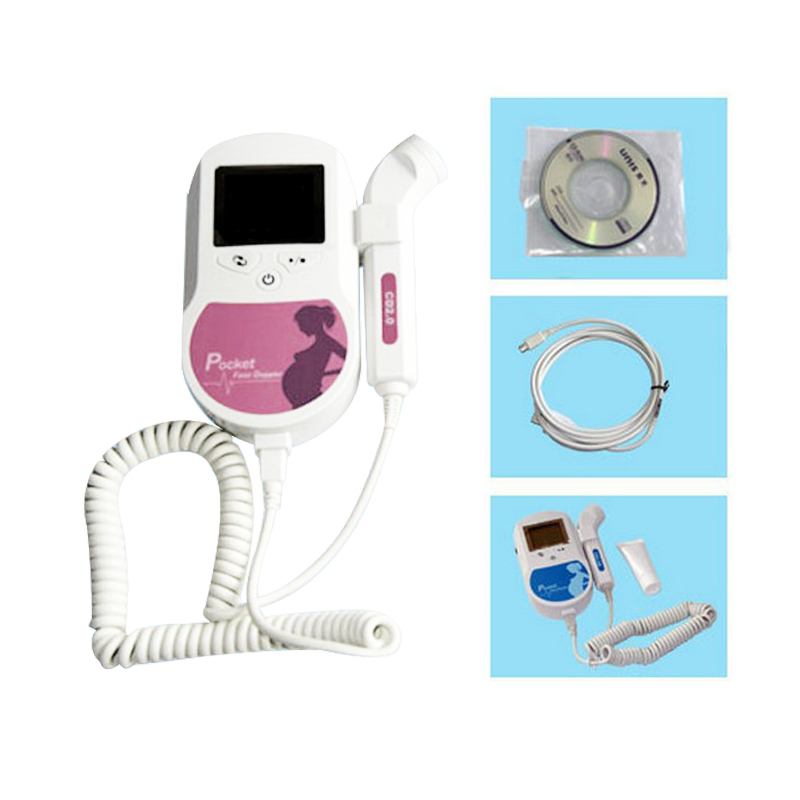
#Doppler ultrasound pregnancy for nclex pro
Students: Educators’ Pro Tips for Tough Topics.Maternity Nursing and Care of the Childbearing Family.


 0 kommentar(er)
